Following on from our post on pregnancy diagnosis, let’s look at using US to assess foetal viability.
Size, movement and presence/absence of Doppler flow can be used to get a general overview but foetal heart rate (FHR) is the most definitive and reliable method of assessing foetal viability.
Using FHR to assess foetal viability:
Foetal HR should be about 1.5-2 times the maternal heart rate but the following is a rough guide:
FHR >180-220bpm – normal
FHR 150-180bpm – foetal stress
FHR<150bpm – C-section recommended
To calculate HRs, use M-Mode or pulsed wave Doppler (PW) and place the vertical callipers between foetal diastolic excursions. You need to know how many cycles your machine uses to calculate HR. (hint: on a Mindray the number of cycles appears as a number in brackets: HR (2) indicates that the cursors be placed over 2 diastolic excursions).
Other sonographic indicators of foetal viability:
Movement – movement indicates viability but Excessive motion indicates foetal stress.
Blood Flow – Presence of colour/power Doppler flow is encouraging but absence of Doppler can be due to inappropriate angle of insonation or machine settings so is unreliable.
Size – Obvious variation in foetal size suggests death +/- resorption of one or more foetus.
Heartbeat – Cardiac activity may be seen from Day 23-25.
FHR – Most reliable indicator of viability is FHR and can normally be measured from Day 28.
Sonographic signs of foetal death:
Absence of heartbeats, variable foetal sizes, poor differentiation of anatomy at expected gestational age, reduced volumes of allantoic and amniotic fluid with increased echogenicity, gestational sacs of varying sizes and shapes if foetal death occurs at different times with different rates of resorption. Death prior to Day 25 usually results in foetal resorption so may see reduction in size and echogenicity of sacs/foetuses as they are resorbed over time. Death after Day 25 usually results in the delivery of mummified or macerated tissue. Subsequent repeated scans may be needed to assess changes over time.
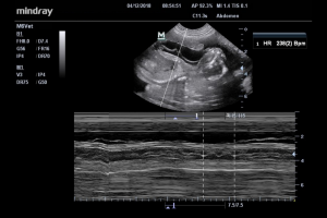
Measurement of FHR using M-Mode on the Mindray M6Vet with the C11-3s probe.
How do you calculate FHR on your ultrasound machine? Download our PDF guide →