In this final part of our introduction to scanning lungs in all species, we discuss the “C” in the ABC’s of lung ultrasound
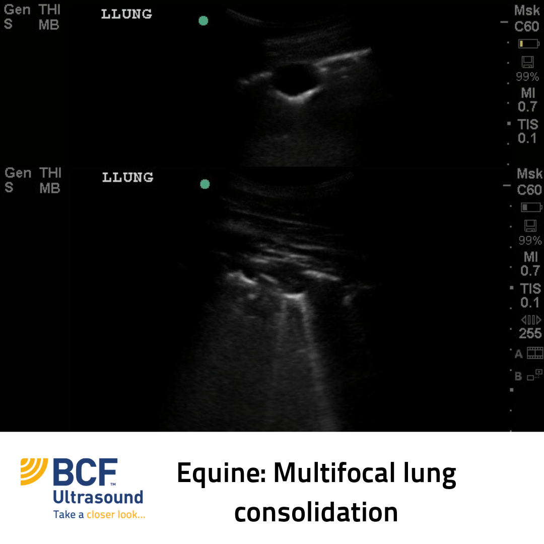
C is for… Consolidation
Lung consolidation occurs when the normally gas-filled pulmonary parenchyma is infiltrated with fluid/cells. When this happens, hypoechoic tissue replaces the normal highly echogenic, linear pleural surface. Lung consolidation is often referred to as ‘hepatised’ lung because the lung develops the ultrasound appearance of normal liver.
 Differential Diagnoses for Consolidation
- Pneumonia (e.g. Rhodococcus equi pneumonia in foals)
- Oedema (severe)
- Lung lobe torsion
- Pulmonary contusions in patients with a history of trauma
- Neoplasia
Consolidation vs. Atelectasis
Atelectasis secondary to pleural effusion is seen readily on ultrasound examination. With atelectasis, the ultrasound appearance is similar to consolidation, but the lung has decreased in volume. Residual alveolar and bronchial air will form multifocal echogenic linear structures (air bronchograms).
Note: Atelectasis secondary to pneumothorax cannot be visualized with ultrasound due to surrounding air interfaces. Use thoracic radiography in these cases.
In the clip below you will notice the similarity in echogenicity and echotexture between this dog’s lung and liver. This dog had pulmonary adenocarcinoma with a highly cellular pleural effusion. In this case, is the lung consolidated or atelectic? Comment below…
References:
Larson MM. Ultrasound of the Thorax (Noncardiac). Vet Clin Small Anim 39 (2009) 733-745.