A is for… A-lines
A-lines are artifacts seen in normal lungs. You will be glad to hear that the ultrasound appearance of the normal lung is the same in all species – human, dog, cat, foal or calf!
When ultrasound beams travel through the chest wall and hit the lung interface, up to 99% of the beams are reflected back. Because these beams still have loads of energy, they keep bouncing back and forth until their energy finally attenuates.
When the beams bounce back the first time, the bright white line you see sliding back and forth underneath the chest wall will be the true pleural surface.
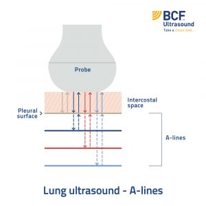
When the beam bounces back to the probe a second time, the machine thinks that the beam has traveled TWICE the distance. The machine “falsely” places a bright white horizontal line an equal distance down the image. And so on for the third time, and fourth time, etc. (see Figure 1).
These parallel white lines equally spaced down the image are your “A-lines”. They are a “useful” artifact because they tell you that this area of the lung surface is normal. The clip below shows you what a normal lung ultrasound looks like…
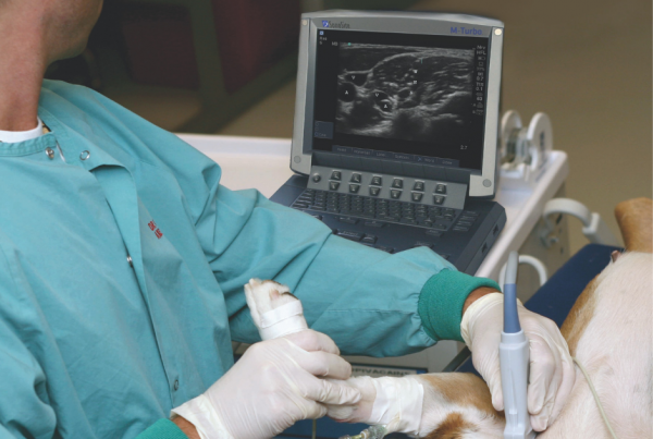
In Part 1 we discussed what probes you need for performing chest scans in your patients.
In Part 3 we will discuss the “B” in The ABCs of Lung Ultrasound.
References:
Noble VE & Nelson BP (2011). “Chapter 1: Fundamentals.” In: Manual of Emergency & Critical Care Ultrasound. Pp: 1-22.